Overview
The Alberta government manages fish diseases of concern in Alberta through a variety of programs. These include:
- monitoring, inspection and detection with lab testing through the Fish Disease Laboratory
- prevention of introduction with regulations and policies
- biosecurity measures such as watercraft inspections and decontamination and the restriction of movement of fish
- sourcing water from a disease-free lake or waterbody and water treatment such as ultraviolet radiation and ozone treatment for fish hatcheries
- public education on how to identify diseases in fish, and how to prevent the spread
- management of diseases in non-infected versus infected zones with decontamination protocols
Diseases of concern
The following fish diseases of major concern are monitored in Alberta and across Canada to ensure early detection, prevent the introduction and spread of diseases and effectively respond, manage and contain outbreaks.
-
Bacterial Gill Disease
Causative agent
Bacterial Gill Disease (BGD) is caused by the bacterium Flavobacterium branchiophilum.
Impacts
BGD is a major concern in freshwater aquaculture facilities. When environmental conditions are favorable, the bacterium attaches and proliferates on gill tissue affecting gill function and leads to high mortality.
Signs and symptoms
The disease is characterized by the presence of large number of filamentous bacteria on the gills. External signs may include lethargy, loss of appetite, clubbed gill filaments, gills appear swollen and protrude from under the operculum, large number of affected fish gather near screen or outlet of pond, swimming high in the water and in a tilted position signalling difficulty in breathing.
Testing
The lab uses microbiological culture technique to detect the causative agent of BGD.
Detections in Alberta
BGD causative agent has been previously isolated from Alberta wild fish.
Control measures
Management strategies such as water quality improvement through treatment with antiseptic and odour control agents have been found to be effective.
Image 5. Rainbow trout affected with bacterial gill disease showing severely swollen gills.
 Source: Dr. Chris Wilson, Utah Division of Wildlife Resources.
 -
Bacterial Kidney Disease
Causative agent
Bacterial Kidney Disease (BKD) is caused by the bacterium Renibacterium salmoninarum and it severely affects the fish kidney.
Impacts
BKD causes high mortality in cultured fish and in wild population. BKD is a systemic bacterial infection that affect juvenile and adult salmonids.
Signs and symptoms
Internally, the distinctive signs of the BKD include focal to multifocal greyish‐white lesions or cysts (granuloma) on the kidney and sometimes in the spleen and liver, ascites in the abdominal cavity, hemorrhages on the abdominal wall or internal organs, swollen kidney, convex, corrugated or lumpy surface of the kidney. External signs may include spawning rash, darkened body, bulging eyes and swollen abdomen.
There is no human health concerns associated with R. salmoninarum.
Testing
Diagnosis of BKD causative agent is done by culture and serology. R. salmoninarum is difficult to culture and requires long incubation period. In addition to culture, PCR and indirect fluorescent antibody technique are commonly used to detect R salmoninarum.
Detections in Alberta
BKD has been previously found in Alberta, but BKD infections is more prevalent in British Columbia in coho salmon, rainbow trout, sockeye salmon, chinook salmon and Atlantic salmon. BKD is a reportable disease in Canada.
Control measures
Management strategies such as broodstock screening, vaccinations and good husbandry practices may reduce its prevalence in hatcheries.
Image 4. Salmon infected with bacterial kidney disease, showing profound greyish-white cysts (granulomas) on kidney and liver.
 Source: An overview on understanding the major bacterial fish diseases in freshwater salmonids. Frontiers in Aquaculture 2025. Imtiaz Ahmed, Shagufta Ishtiyaq and Shabihul Fatma Sayed. DOI:10.3389/faquc.2025.1515831.
 -
Ceratomyxosis
Causative agent
Ceratomyxosis disease is caused by the parasite Ceratomyxa shasta and it affects salmon and trout.
Impacts
Ceratomyxosis causes mortality in young finfish in hatcheries and previously infected adult finfish returning to spawn.
Signs and symptoms
The parasite is mainly found in the intestine but can also be found on the gills (actinospores). Signs of the parasitic infection may include darkened skin colour, an enlarged belly filled with fluid, pus in the liver and spleen, protruding or reddened vent, bulging eye, bleeding in the intestine and internal organs.
Ceratomyxa shasta does not pose a risk to human health.
Testing
Diagnosing ceratomyxosis requires laboratory testing. Ceratomyxosis is a federally reportable disease in Canada and as such any suspect cases must be reported to the Canadian Food Inspection Agency.
Detections in Alberta
This parasite has not been found in Alberta but has been found in wild finfish in the Pacific Ocean watershed of British Columbia.
Control measures
Preventive measures such as restrictions of movement of fish, strict fish import licensing by regulatory authorities, proper disposal of fish wastes, proper washing and disinfecting of footwear and fish gear and strict biosecurity rules help to prevent the introduction and spread of this parasite.
-
Columnaris disease
Causative agent
Flavobacterium columnare is the causative agent of Columnaris disease in cultured and wild freshwater fish species such as salmonids, carp, catfish, goldfish, perch and tilapia.
Impacts
Columnaris disease may cause mass die-off of wild fish population and high mortality in cultured fish.
Signs and symptoms
The characteristic external signs of this disease may be an appearance of a saddle shaped white area around the dorsal fin, gill necrosis and erosion and ulceration of the skin. It could also manifest in patchy skin and fin discolouration with worsening erosion and eventual sub cutaneous deep ulceration. Skin lesions on infected fish typically originate at the base of the dorsal fin and extend ventrally on either side of the body forming the classic saddle back.
Testing
The lab screens cultured and wild fish samples for F. columnaris using bacterial culture media. Wet mounts of skin and gill lesions may show filamentous bacteria with gliding motility forming a haystack-like arrangement.
Detections in Alberta
Mortalities from Columnaris infection has been reported in white sucker, yellow perch and white fish in Saskatchewan lakes but some F. columnaris isolates have been isolated from fish of Alberta origin.
Control measures
Water quality management, the use of antibiotics and biosecurity measures are effective in preventing the introduction of the bacterium into a hatchery system.
Image 7. Rainbow trout with Columnaris disease, showing cotton wool-like growth (left photo) and characteristic ulceration along the lateral body wall (right photo).
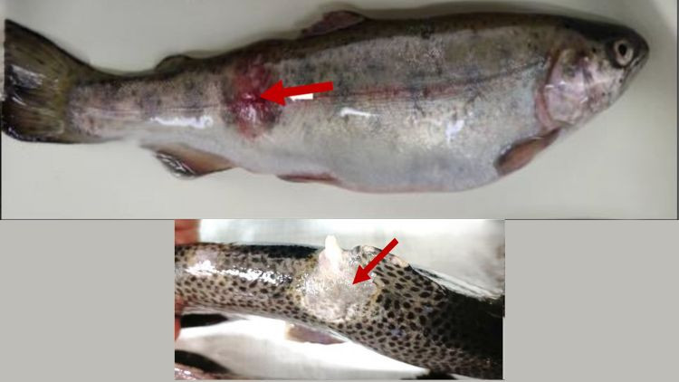 Source: An overview on understanding the major bacterial fish diseases in freshwater salmonids. Frontiers in Aquaculture 2025. Imtiaz Ahmed, Shagufta Ishtiyaq and Shabihul Fatma Sayed. DOI:10.3389/faquc.2025.1515831.
 -
Enteric Redmouth disease
Causative agent
Enteric Redmouth disease (ERM) is a bacterial disease in salmonids caused by the bacterium Yersinia ruckeri.
Impacts
This disease affects salmonids in fresh water, particularly rainbow trout where it causes high mortality in fry and fingerlings. ERM has been reported in species such as whitefish, steelhead trout, cutthroat trout, chinook salmon, lake trout, burbot and Dolly Varden trout.
Signs and symptoms
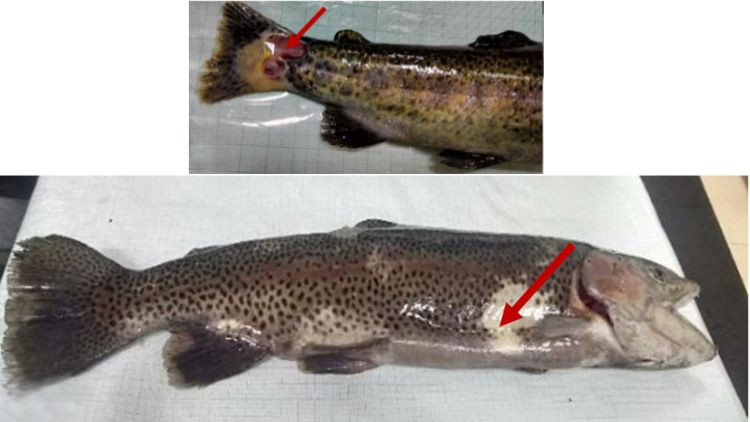
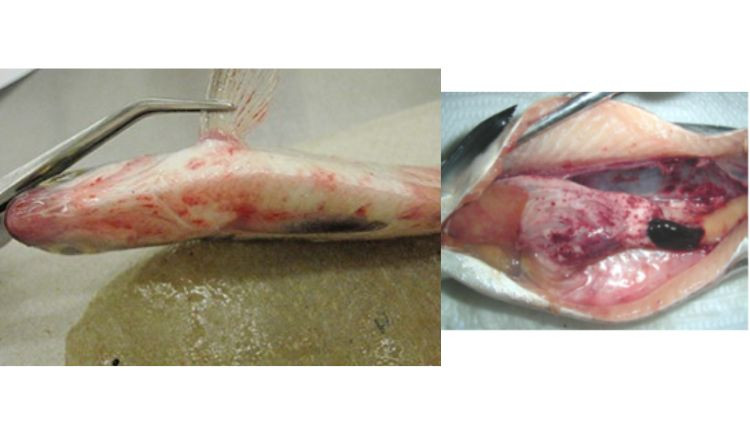
This disease is characterized by subcutaneous hemorrhages at the corners of the mouth, jaw and in the gums and tongue, hemorrhages on the surface of the liver, pancreas, pyloric caeca and swim bladder. Other signs may include darkening of the skin, inflammation of the lower intestine, enlarged spleen, lethargy, loss of appetite and swimming at the surface of the water.
Testing
The lab tests for ERM causative agent using microbiological culture method. Suspect cases of ERM must be reported to the Canadian Food Inspection Agency for proper actions and response.
Detections in Alberta
ERM has been previously found in Alberta and other regions of Canada.
Control measures
In cultured fish, ERM infections can be managed by good husbandry practices such as cleaning and disinfecting of equipment, use of footbaths, water quality management, vaccination and good stock management.
Image 8. Rainbow trout showing clinical signs of enteric redmouth disease. Hemorrhages in the caudal and anal fins (arrows), enlarged and black spleen and reddened intestine. Note: S: spleen, K: kidney and I: Intestine.
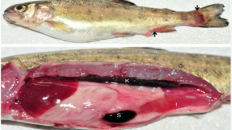 Source: Kumar, G., Hummel, K., Noebauer, K. et al. 2018. Proteome analysis reveals a role of rainbow trout lymphoid organs during Yersinia ruckeri infection process. Sci Rep 8, 13998 (2018). https://doi.org/10.1038/s41598-018-31982-6.
 -
Furunculosis
Causative agent
Furunculosis is a bacterial disease primarily affecting wild and cultured salmonids and other freshwater fish caused by the bacterium Aeromonas salmonicida.
Impacts
Furunculosis causes mortality in wild and cultured salmonids, leading to significant economic losses in hatchery operations.
Signs and symptoms
Acute cases appear as generalized bacterial septicemia characterized by external hemorrhagic lesions at the base of the fins and oral cavity, dark body colouration, lack of appetite, erratic swimming, lethargy and high mortalities, within few days.
Testing
Furunculosis is a reportable disease of finfish in Canada. The lab routinely screens wild and hatchery samples for the presence or absence of Aeromonas salmonicida using bacterial culture method.
Detections in Alberta
This disease has been reported in Alberta and other parts of Canada.
Control measures
Effective implementation of disease surveillance and biosecurity measures can prevent the spread or introduction of the disease.
Image 6. Rainbow trout with furunculosis, showing deep ulcerative skin lesions (left side photo) and profound body discolouration (right side photo).
 Source: An overview on understanding the major bacterial fish diseases in freshwater salmonids. Frontiers in Aquaculture 2025. Imtiaz Ahmed, Shagufta Ishtiyaq and Shabihul Fatma Sayed. DOI:10.3389/faquc.2025.1515831
 -
Infectious Hematopoietic Necrosis disease
Causative agent
Infectious Hematopoietic Necrosis virus (IHNV)
Impacts
Infectious Hematopoietic Necrosis (IHN) is commonly called “pop-eye disease” and can cause significant mortality in young, farmed trout and salmon fry, fingerlings or adults during or after spawning.
Signs and symptoms
IHN infection is characterized with darkening of the fish body from the tail region, swollen abdomen, swelling of the kidney, exophthalmia (pop-eye), hemorrhaging at the base of fins, around the eye, pale gills, lethargy and abnormal or erratic swimming behaviour. White to yellowish fluid may be present in stomach and intestines.
Testing
The lab plays a significant role as part of Alberta’s fish health regulator in testing and screening for the IHNV. IHN is a reportable disease in Canada, implying suspect cases must be reported to the Canadian Food Inspection Agency.
Detections in Alberta
Alberta is considered free of IHNV. The IHNV has been found in wild finfish in the Pacific Ocean watershed of British Columbia. There are no known treatments for IHN, but vaccines have been effective against the IHN virus.
Control measures
IHN can be spread by moving infected live or dead finfish, contaminated equipment or contaminated water. Biosecurity measures such as import permit or license required before moving fish, fish movements restrictions, proper cleaning and disinfection of fish gears and footwear after working with finfish in the wild are targeted measures to prevent the introduction and spread of infectious hematopoietic necrosis virus.
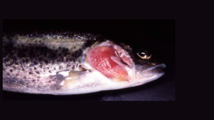
Image 2. Signs of IHNV disease in rainbow trout include hemorrhage around the eye and exophthalmia (pop-eye – left photo), and skin darkening relative to lighter coloured healthy fish (right photo).
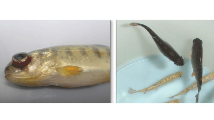 Source: Gael Kurath, Western Fisheries Research Center, U.S. Geological Survey, Seattle. WA.
 -
Infectious pancreatic necrosis disease
Causative agent
Infectious pancreatic necrosis virus (IPNV)
Impacts
Infectious pancreatic necrosis (IPN) causes high mortality in fry and fingerlings of salmon and trout. It is highly virulent in younger fish of less than 6 months of age, while older fish are less susceptible. It is a disease of great concern in aquaculture.
Signs and symptoms
In hatchery populations, the first sign of the disease is a sudden increase in daily mortalities in fry and fingerlings. External signs of the disease may include darkening of the skin, abdominal swelling, abnormal spiralling swimming motion and bulging of the eyes. Internally, infected fish may have hemorrhage over the anterior visceral mass, enlarged, pale liver and spleen. A distinct characteristic of IPN infection is the presence of large amount of whitish or milky mucus in the stomach and intestine.
Testing
The lab routinely screens Alberta’s wild and hatchery samples for IPN virus using cell culture and real time polymerase chain reaction (PCR). It is a reportable disease in Canada and any suspect case must be reported to the Canadian Food Inspection Agency.
Detections in Alberta
In 1984, IPNV was detected in wild lake trout population of northern Alberta through routine testing.
Control measures
IPN control is difficult in the wild as it is spread through any excrement from infected fish, fish to fish and the water they live in. In hatcheries sourcing their water from streams containing the IPN virus, ozone treatment of the water is effective to eliminate the IPN virus. Control measures have been implemented to prevent its spread to non-infected lakes through biosecurity and reporting of suspected cases.
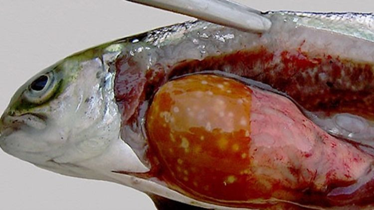
Image 1. Atlantic salmons infected with infectious pancreatic necrosis disease showing diffuse hemorrhages in visceral fat, pyloric caeca and yellow or pale liver.
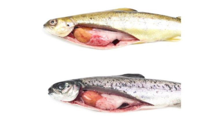 Source: Godoy et al., 2022. Isolation of a New Infectious Pancreatic Necrosis Virus (IPNV) Variant from Genetically Resistant Farmed Atlantic Salmon (Salmo salar) during 2021–2022. Pathogens 2022, 11(11), 1368; https://doi.org/10.3390/pathogens11111368
 -
Proliferative Kidney Disease
Causative agent
Proliferative Kidney Disease (PKD) is a parasitic disease of wild and farmed salmonids, particularly rainbow trout and it’s caused by the parasite, Tetracapsuloides bryosalmonae.
Impacts
PKD may cause mass die-off of wild fish population. The disease is temperature-dependent, with outbreaks often occurring in warmer summer months. Proliferative kidney disease is caused by pre-sporogonic stages of the parasite that infect the kidney interstitium and other vascularized organs.
Signs and symptoms
Internal signs of the disease include enlargement of the spleen and the posterior or entire kidney, grey or mottled kidney, distended kidney capsule and ascites. Fish with PKD may appear lethargic and dark, with distended abdomen, pale gills and bulging eyes.
Testing
The lab routinely screens salmonids for T. bryosalmonae using qPCR.
Detections in Alberta
PKD has been detected in Alberta, and is currently being monitored to determine the distribution and impacts on fish populations
Control measures
Monitoring, surveillance and biosecurity measures are effective in preventing the introduction or spread of PKD. Clean, Drain, Dry and decontamination protocols should always be followed to reduce the spread of the parasite.
Image 10. Proliferative Kidney Disease in rainbow trout exhibiting bloody ascites and swollen nodular posterior kidney due to inflammation.
 Source: M. L. Kent and R. P. Hedrick, University of California, Davis.
 -
Viral Hemorrhagic Septicemia disease
Causative agent
Viral Hemorrhagic Septicemia virus (VHSV)
Impacts
Viral Hemorrhagic Septicemia (VHS) may cause mortality in cultured fish but more prevalent in wild fish from fresh water and marine environments, causing mass fish die-offs affecting several fish species such as walleye, yellow perch, Northern pike, muskellunge, largemouth and whitefish. Mortality is highest at lower water temperatures, from 2 to 15 C, and during spawning. VHS may affect fish of all ages and sizes. Salmonids (trout and salmon) are less susceptible to the virus.
Signs and symptoms
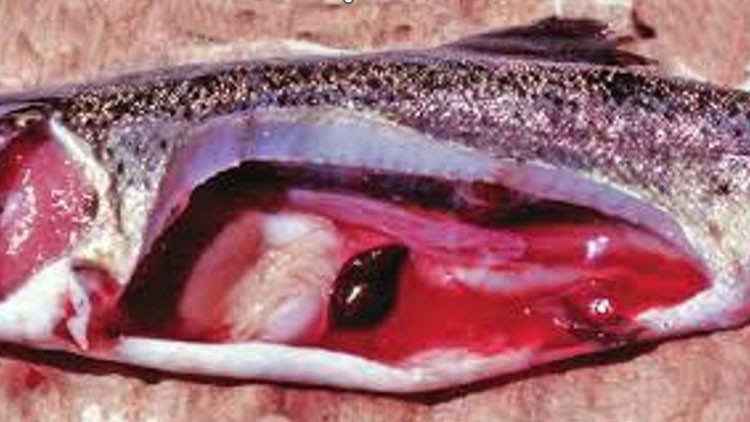
Signs of VHS disease include hemorrhage in the skin, base of fins, eyes and gills, pale gills and organs, bloated abdomen, bulging eyes, gasping at the surface, darker body colouration, multifocal hemorrhages on liver and skeletal muscles, diffused hemorrhages on viscera, swollen kidney and liver.
VHS has no known risk on human health. However, fish that appear ill, dying or that are found dead should not be consumed by the public or anglers.
Testing
Alberta’s wild and hatchery-cultured fish are routinely screened for VHS using cell culture and PCR techniques.
Detections in Alberta
To date, no cases of VHS has been reported in Alberta, but the VHS virus has been detected in the Great Lakes watershed of Ontario, Nova Scotia, New Brunswick and US bordering states.
Control measures
VHS may spread from one waterbody to another through the movement of fish, water, vessels and equipment that have come in contact with the virus. Measures such as safe disposal of fish waste, fish movement restrictions, public education, routine sampling, disease screening and testing prior to fish movement, proper cleaning and disinfection of fishing gears, trailers and footwears by anglers and boaters are effective ways to prevent the spread of VHS virus.
Image 3. Left photo – Fish with external signs of viral hemorrhagic septicemia – extensive hemorrhage on the skin, base of fins, eyes and gills. Right photo – Extensive diffused hemorrhages on viscera, liver and skeletal muscles.
 Source: Left photo – Dr Thomas Loch, Michigan State University (MSU-AAHL) Aquatic Animal Disease Ecology Program. Right photo – British Columbia Centre for Aquatic Health Sciences, Campbell River.
 -
Whirling disease
Causative agent
Whirling disease is caused by the myxozoan parasite Myxobolus cerebralis.
Impacts
This parasite infects wild and cultured salmonid populations and can cause high mortality in trout populations.
Signs and symptoms
Infected fish may have deformities of the head, body, or tail. The tail may also be darkened, sometimes even looking black. Infected fish may be seen swimming in circles (whirling movement).
Testing
Whirling disease is a reportable disease in Canada. The lab tests for the causative agent of whirling disease, M. cerebralis, by performing qPCR using pepsin-trypsin digested cartilage or homogenized cranial tissues from fresh or frozen fish as the substrates. The lab has been a major hub for extensive whirling disease testing since 2018 in the province of Alberta.
Detections in Alberta
In Canada, the disease was first detected in fish from Johnson Lake, Banff National Park, Alberta in 2016. This resulted in a robust outbreak response, surveillance and monitoring through Alberta’s whirling disease program. Whirling disease was detected in Yoho National Park in 2024 and Kootenay lake in British Columbia in 2025.
Control measures
There is no treatment for whirling disease. Biosecurity measures are effective in preventing its spread by following decontamination protocols and making sure to always clean, drain and dry all equipment that contacts the water.
- Learn more about whirling disease
Image 9. Rainbow trout infected with whirling disease showing cranial and spinal deformities, crooked and blackened tail.
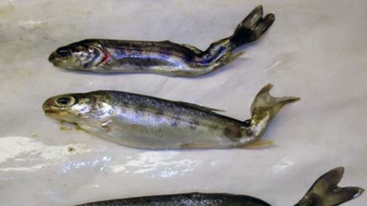 Source: Sascha Hallet and FishPathogens.net